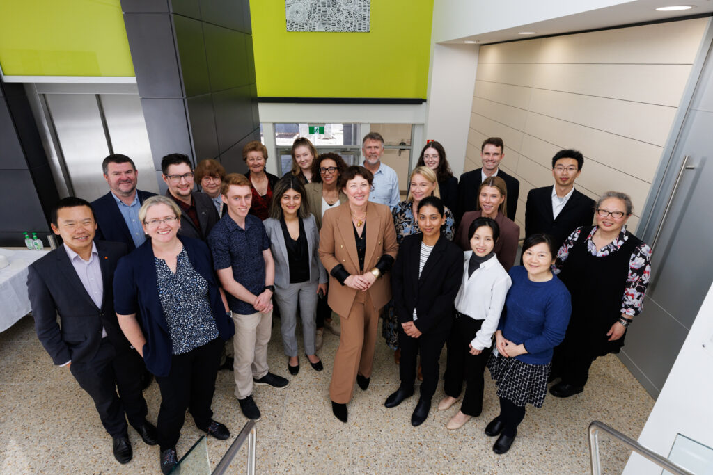
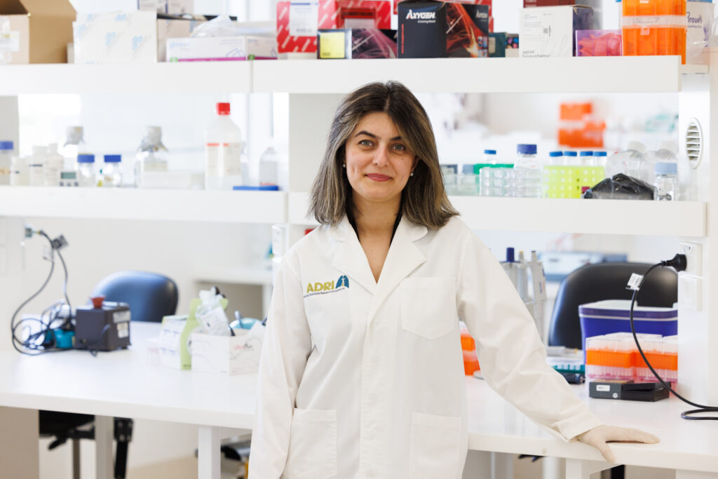
ADDRI is an independent, not-for-profit research institute committed to reducing and ultimately eliminating the impact of asbestos and dust-related diseases worldwide.
Our team of doctors, researchers, scientists, nurses and industry leaders excel in innovative thinking and strive for excellence every day.
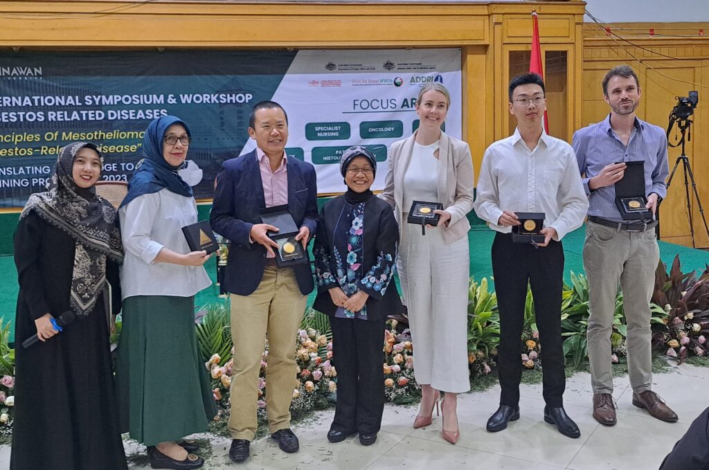
The importance of collaboration
We are on a mission to collaborate with those working toward the elimination of asbestos and dust-related diseases.
About the Diseases
Asbestos and dust diseases remain a clear and present danger. 4,000 Australians die each year from asbestos-related diseases. It is estimated that one in four workers are at risk of silica-related disease.
Asbestos is a type of mineral made up of tiny, needle-like fibers. Known for its ability to resist heat, electricity, and corrosion, it was widely used in many construction materials. Inhalation of asbestos fibres can directly lead to disease and a form of cancer called mesothelioma.
What is Silica?
Crystalline silica is a natural mineral found in sand, stone, concrete and mortar. Found in high quantities in materials commonly used for tunnel construction and engineered stone, inhalation of silica dust particles can cause serious disease.
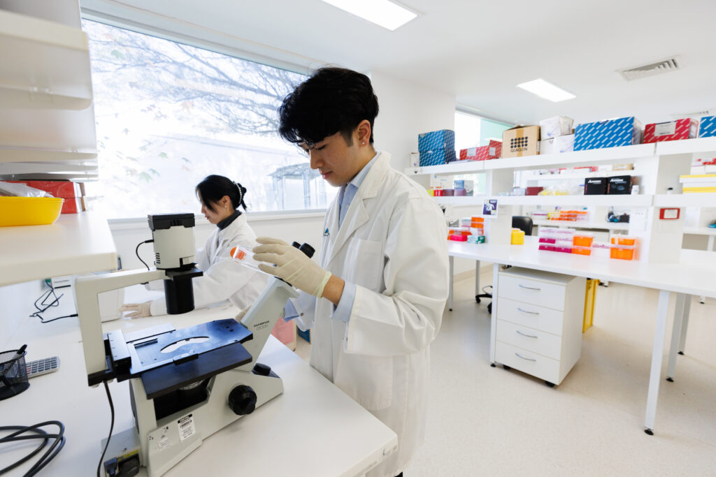
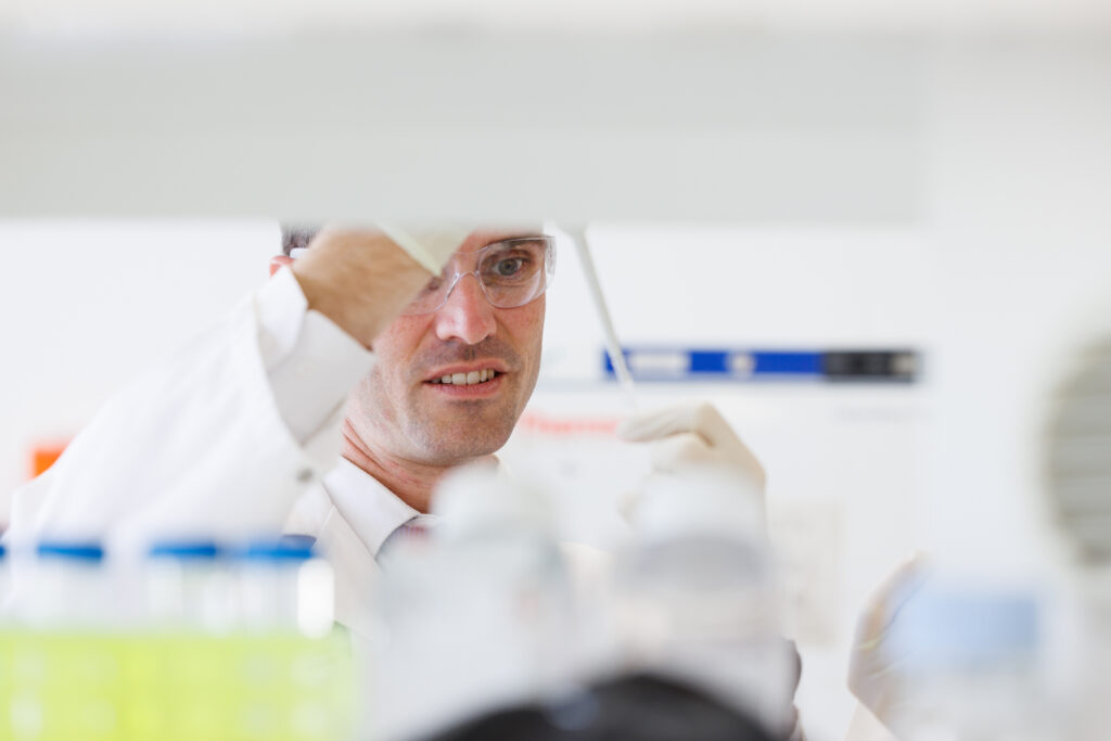
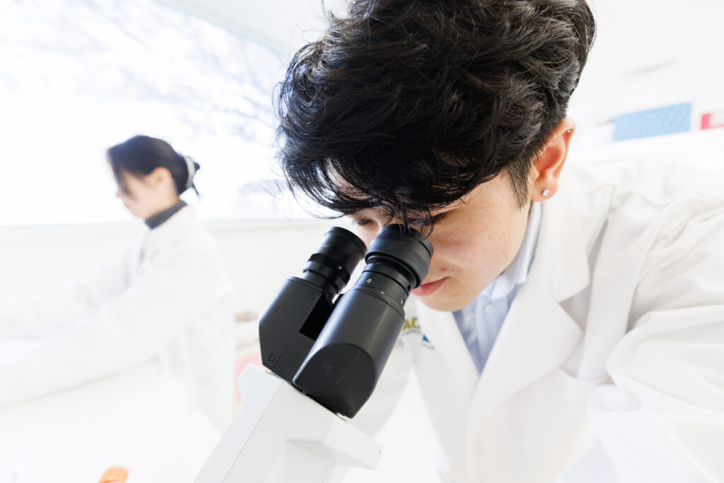
Research
We are on a mission to understand asbestos and dust-related disease development and diagnosis through dedicated research. Our evidence-led research drives advancements in medical and scientific understanding and guides our global education and training initiatives.
We are unrelenting in exploring every avenue that will achieve our mission of improving patient outcomes, uncovering new treatments and ultimately ending all asbestos and dust-related diseases.
Working globally
Our status as the WHO Collaborating Centre for Elimination of Asbestos-Related Diseases ensures that our dedication, research and collaboration will have significant impact around the world.
Support
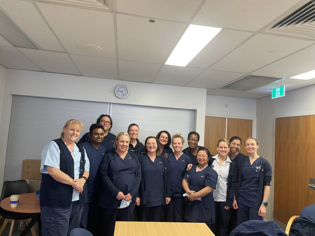
We are on a mission to support people impacted by dust-related diseases. Our Mesothelioma and Silicosis Support Service addresses the needs of patients suffering dust-related diseases and supports their families.
Our specialist nurses are here to address the needs of our patients with mesothelioma and to support carers and families. We can answer any questions you may have and help you navigate the health system
Silicosis support
Emotional and practical support for individuals diagnosed with silicosis and their caregivers.
Education
We are on a mission to educate that asbestos is a very real threat to people all over the world and provide the knowledge and tools to care for those impacted.
An internationally recognised eToolkit on asbestos-related diseases (ARDs), important research to fill knowledge gaps on the elimination of asbestos and much needed training on ARDs in developing countries.
Mesothelioma learning module
Access online training for nurses/health care professionals to assist with diagnosing and caring for patients with mesothelioma.
Mesothelioma is a peculiar type of malignancy, which is highly related to asbestos-exposure, because 80 to 90% of patients with mesothelioma have a history of occupational and/or environmental exposure to asbestos.
Mesothelioma is located in the pleura, the peritoneum, the pericardium and the tunica vaginalis, all of which is normally lined by mesothelial cells. The population of each of the site is 70-80% in the pleura, 10-20% in the peritoneum and several percent in the pericardium and the tunica vaginalis.
Diffuse spread along the cavity is characteristic in most of mesothelioma, but rarely (probably a few percent) it forms a localized tumor without the diffuse spread pattern.
Mesothelial cell primarily has a potential of differentiation to an epithelioid cell lined at the serosal surface as well as a mesenchymal spindle-shaped cell presented under the serosal lining. Therefore, mesothelioma has various histological subtypes as shown Table 1, including carcinoma-like (epithelioid) tumor, sarcoma-like tumor (sarcomatoid) tumor, or mixed (biphasic) tumor with both components (epithelioid and sarcomatoid).(1)
Desmoplastic mesothelioma is basically sarcomatoid mesothelioma, but in more than 50% of the tumor area, abundant proliferation of collagen fibers is seen, showing storiform pattern or with no pattern. Also, many histological rare variants are known. Lymphohistiocytoid mesothelioma consists of large histiocytoid cells with some features of mesothelial cells, associated with lymphocytic infiltration. Deciduoid mesothelioma consists of large cells with abundant weakly-eosinophilic cytoplasm and centrally located round nucleus like decidual cells. Mesothelioma with heterologous elements is defined as a tumor including the findings of osteosarcoma, chondrosarcoma or rhabdomyosarcoma.
Two mesothelial tumors with benign behavior are known. Well-differentiated papillary mesothelioma (WDPM) shows papillary structure with fibrovascular core.(2) The lining cells are well-differentiated with no significant atypia. WDPM is originally reported in the pleura, but occasionally peritoneal cases are reported. Adenomatoid tumor originates in the uterus, but rarely occurs in pleura and peritoneum. The tumor consists of small tubules with fibrous stroma. The lining cells have no significant atypia. These two types of mesothelial cell tumor are necessary to differentiate from epithelioid mesothelioma.
The proportion of histological types is: about 60% epithelioid mesothelioma, about 20% sarcomatoid mesothelioma (including desmoplastic mesothelioma), about 20% biphasic mesothelioma. Other types are rarely seen.
Differential Diagnosis
It is important to remember that there are many diseases to be differentiated when mesothelioma is diagnosed, as shown in Table 2.(3)
In the pleura, the invasion of adenocarcinoma of the lung with collagenous stroma is common. Even if the primary carcinoma is small beneath the visceral pleura, wide extensive growth occurs along the pleura, which is called “pseudo mesotheliomatous adenocarcinoma.” Squamous cell carcinoma appears in the same manner. In the lung, carcinoma with sarcomatoid features, which is called “sarcomatoid carcinoma” or “pleomorphic carcinoma” is well known. The differentiation should be based on the observation of H&E specimen, however, immunohistochemical (IHC) staining is very useful. (4,5) (Table 3)
In the peritoneum, ovarian or peritoneal serous papillary carcinoma is difficult to be differentiated. On the basis of knowledge in embryology, the nature of ovarian surface cell is similar to that of mesothelial cell, with common origin of coelomic epithelium and therefore IHC staining is necessary to ascertain phenotype of tumor cells. (Table 4)
Various histological types of sarcoma have to be differentiated from sarcomatoid mesothelioma in the pleura and the peritoneum.(6) In the peritoneum, sarcomatoid mesothelioma is very rare. (Table 5)
For the decision of treatment, the differentiation between benign lesion (fibrous pleuritis) and sarcomatoid mesothelioma is very important. In the pleura, misdiagnosis of sarcomatoid or desmoplastic mesothelioma occurs very often. Especially the histological examination of small pieces of pleural tissue should be diagnosed very carefully. The attending clinician should submit an abundant and perpendicularly-deep specimen to the pathologist. (Table 6)
The differentiation between reactive mesothelial cell proliferation and epithelioid mesothelioma in situ (or of early stage) is also difficult.(7,8) Careful observation of the HE specimen is necessary, including IHC stainings. Finally, genetic alteration should be examined by means of FISH. (9,10)(Table 7)
Telepathology
The remote system of pathological diagnosis is very different from the traditional method of utilizing a microscope. This new advanced method of pathological diagnosis is expected to become a new standard for future generations of health professionals. There may be some hurdles to adopting this system, such as budget limitations which may make purchasing these instruments difficult. There are, however merits relative to the traditional method.
Necessary items
Whole slide scanner: an instrument to scan a glass-slide and create a digital image
LOOKREC®: a cloud-based system for remote diagnosis which has been developed by MNES Inc.
Actual process
A glass-slide is prepared with various stains done in the same way as traditional methods.
Scanning is done by the whole slide scanner. Set the glass-slides in the scanner and locate the area to scan as well as which point to focus on. Several minutes are necessary to complete the scan.
Upload the digital image to the cloud storage of LOOKREC®. Once it has been uploaded to the cloud, you will be able to access the image database and make a diagnosis.
Advantages of digital pathology
When utilizing the traditional method, there is the problem of storing glass-slides as they are heavy and take up much space in the laboratory. In comparison, digital images do not take up physical space in the lab.
The color of staining glass-slides fades over time. The digital images do not fade or change as the data is stored in the cloud.
Making comparative observations between the previous and current data is much easier. The previous glass-slide data is stored according to the patient ID number which comprises a large patient database.
If special staining or immunohistochemical (IHC) staining is used for making a diagnosis, one monitor image can be divided into several smaller images.
Education, training, and research
When the advantages of digital pathology are considered, a new frontier of pathological diagnosis can be imagined.
An international consultation system of pathological diagnosis using digital pathology and LOOKREC® can be introduced. At present, a consultation network is functioning between Mongolian pathologists and Japanese pathologists . Without such a system, the glass-slides would have to be sent carefully by post. It would be impossible to make a diagnosis in a timely fashion under these conditions.
In many Asian developing countries, many doctors are still in early stages to develop understanding of ARDs. Although asbestos is commonly used in Asia, ARDs are still largely under-diagnosed. Therefore, a specific database of ARD should be established by a dedicated project. Using this database, many medical doctors and medical students will be able to learn from the data in the affected countries.
International collaborative research can be done using digital pathology For example, during the Iran-Iraq war in the nineteen-eighties, mustard gas was used and there were many victims who were exposed in Iran. Japanese and Iranian doctors collaborated on the research to conduct diagnosis and treatment by visiting each other several times. Telepathology would have allowed health professionals to collaborate on diagnosis remotely in a more time and resource-efficient manner.